Dr. Vivek Kumar is the pioneer Coronary Angiography Doctor in South Delhi and provides the latest Coronary Angiography Treatment in Delhi.
Angiography - What is it?
Angiography is a medical procedure that examines the flow of blood in the body. This allows us to intervene and treat obstructions and other conditions, particularly those affecting the heart and brain. The X-ray imaging of blood flow within the body is known as angiography.. Chemicals that are opaque to X-rays are injected into a bloodstream during this medical procedure. It is simple to follow the photos of their blood vessel path. Locating blockages in the heart (coronary), brain (cerebral), lung (pulmonary), and other smaller blood arteries is beneficial.
It can detect abnormal blood flow caused by blood vessel narrowing (known as stenosis), heart structure problems, internal bleeding, or other impediments that should be removed. It may also be useful in locating areas of internal bleeding known as haemorrhage and aneurysms (abnormal dilation of blood vessels). Abnormal blood flow can have an impact on the organs nourished by the vessels, raising the risk of chest pain (angina), heart attacks, strokes, and other illnesses.
Dr. Vivek Kumar is the best Coronary Angiography doctor in South Delhi and is capable of handling even the most difficult cases.
Coronary angiography treatment
Types of Angiography
Coronary Angiography
The coronary arteries supply blood flow to the heart, and if these vessels narrow or block, heart testing may be abnormal, as well as specific symptoms such as:
- Chest pain (angina)
- Change in heart rate
- Change in blood pressure
- Pain
- Congenital heart defecty
- Aortic stenosis
- Heart valve disease
- Chest injury
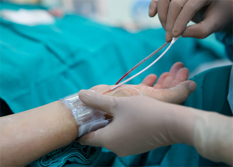
In coronary angiography, a local anesthetic is used to numb the area where the catheter is inserted, which is typically the brachial artery in the forearm or the femoral artery in the groyne. A catheter and guide wire are inserted into the arterial system and checked until they reach the main coronary arteries. The catheter can be moved to image different parts of the arterial system or directly image the inside of the heart. It employs a specialised X-ray dye.
Cerebral Angiography
It may be used to treat narrowing that causes transient ischemic attacks. A clot may be removed and symptoms such as weakness, numbness, vision changes, or loss of speech reversed. It can also be used to seal off cerebral aneurysms, blood vessel bulging, or abnormal dilation that is prone to rupture and secondary haemorrhage.
Microangiography
It could be used to locate the smaller blood vessels that supply other organs.
Preparation for the Coronary Angiography/angiogram
6-hour fasting is required before the procedure.
Few medications must be stopped for 2-3 days prior to the procedure, and others may be continued if you’re Cardiologist so directs.
IIf you are taking insulin to reduce a high level of sugar, you should stop taking it before 1 day to avoid having your sugar level reduced during the procedure
During the Procedure
- Coronary angiography is performed in a cath lab and usually does not require sedation; instead, a local anaesthesia is administered to the site of the wrist or groyne.
- A small catheter is inserted into the artery from the cut side of the wrist or groyne, allowing the catheter to reach the heart.
- An X-ray dye is injected into the coronary arteries via the catheter to look for any blockages that are narrowing the arteries.
- Several X-ray images are now being taken to assess the blockages.
After the Procedure
- After removing the Catheter, pressure is applied to the cut where it was inserted to stop the bleeding.
- In general, the total time required is less than one hour.. After this shifting to the recovery room to rest.
- For a few days, there will be some soreness or bruising at the site of the cut.
- If you feel at ease, you can return home after a few hours.
Risks and Contraindications
- Canesthetic Reactions: Skin irritation at the injection site or fainting may happen.
- Contrast Media: A variety of contrast media are now available, which reduces sensitivity reactions and may result in chemotoxicity and anaphylaxis. Chemotoxicity can occur as a result of contrast media interaction with blood. Side effects include:
- Low blood pressure (hypotension)
- Bradycardia
- Pulmonary congestion
- Contrast-induced nephropathy (CIN)
- Warmth
- Pain, Tightness
- Nausea, Vomiting
- Heparin-Induced Thrombocytopenia (HIT): Heparin is a blood thinner used during angiography. Heparin can cause an amplified immune system response in a few people, activating platelets and causing clotting in blood vessels.
- Local Vascular Injury: There is a risk of bleeding due to blood vessel damage when the catheter is inserted and moved internally.
- Hematoma: When the catheter is removed at the end of the procedure, blood can pool outside of the artery at the site of insertion, forming a mass.called a hematoma.
- False Aneurysm: A false aneurysm (also known as a pseudo aneurysm) can develop when a smaller artery is accidentally catheterized. An incorrect size match can cause damage to the blood vessel wall and the formation of an aneurysm.
- Arteriovenous Fistula (AVF): Arteriovenous Fistula (AVF): It may form when an artery and vein are penetrated near each other and form a connection, which allows the higher arterial pressure to enter the vein.
- Dissection
- Thrombosis and Embolism: A clot, or thrombus, may form; this risk can be reduced by flushing the sheath on a regular basis and using anti-coagulants during longer processes. An embolism is a blood clot that moves through the bloodstream and can cause damage to another part of the body. It can cause a stroke.
- Cholesterol Emboli: An embolism can occur when physical broken cholesterol is deposited along the lining of blood vessels.
- Bradycardia & Tachycardia: Bradycardia (low heart rate) and Tachycardia (high heart rate) can be caused by catheter irritation or blockage as it approaches the heart.